
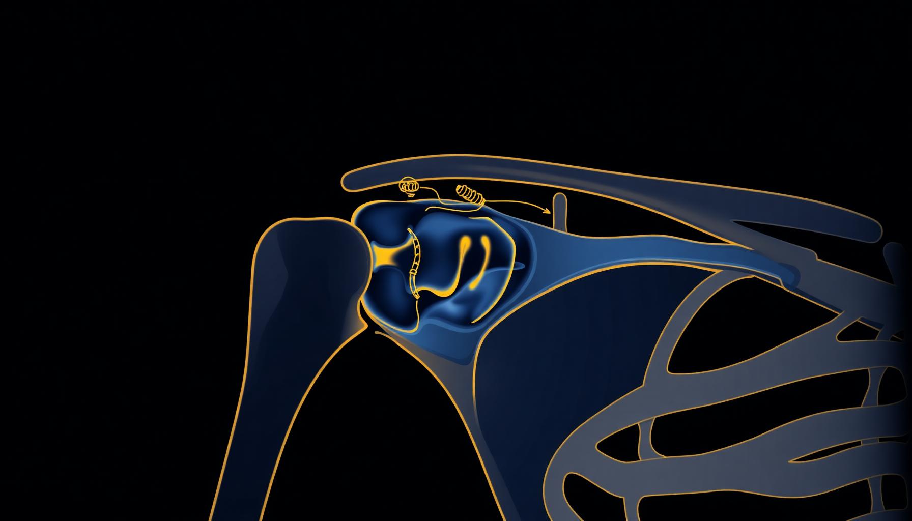
Shoulder Surgeon in Rome
Anatomic and reverse prosthetic surgery, rotator cuff arthroscopy, treatment of shoulder instability.
The most mobile joint of the human body
The shoulder is the most mobile joint in the human body: this extraordinary freedom of movement is made possible by a delicate balance between bone, tendon, capsular and muscular components. This complexity makes the shoulder particularly exposed to degenerative disease, tendon injuries and instability.
Prof. Simone Cerciello treats the full range of shoulder pathology with a personalised approach: from latest-generation anatomic and reverse prosthetic surgery, to minimally invasive arthroscopic surgery for rotator cuff tears and gleno-humeral instability. He is in charge of shoulder and arthroscopic surgery at the A. Gemelli University Hospital IRCCS in Rome.
Shoulder replacement
Joint replacement with anatomic or reverse implants, depending on the pathology and the status of the rotator cuff.
Shoulder replacement is the surgical solution for advanced forms of gleno-humeral osteoarthritis, sequelae of complex proximal humerus fractures and conditions in which the rotator cuff is no longer functional (cuff tear arthropathy).
Two large families of shoulder prostheses exist, profoundly different in biomechanical concept and indications: the anatomic prosthesis, which reproduces the physiological geometry of the joint, and the reverse prosthesis, which inverts the joint geometry to exploit the deltoid muscle as the motor of movement. The choice between the two is defined according to age, bone quality, status of the rotator cuff and functional expectations.
Anatomic prosthesis
What it is
The anatomic shoulder prosthesis faithfully reproduces the natural anatomy of the gleno-humeral joint. The humeral head, damaged by osteoarthritis or other conditions, is replaced with a spherical metal component (chromium-cobalt or titanium alloy), while the glenoid surface is resurfaced with a highly cross-linked polyethylene insert. When wear affects only the humerus (with the glenoid still intact), a hemiarthroplasty replacing only the humeral component may be indicated.
When to operate
The main indication is primary gleno-humeral osteoarthritis, with disabling pain and functional limitation resistant to conservative treatment. Other indications:
- Avascular necrosis of the humeral head
- Sequelae of proximal humerus fractures
- Inflammatory arthritis (rheumatoid arthritis)
- Post-traumatic arthropathies
Essential requirement
An intact and functional rotator cuff. The cuff is in fact the main dynamic stabiliser of the shoulder, indispensable for the correct function of an anatomic prosthesis.
Advantages
- Reproduction of the natural joint biomechanics
- Wide, physiological range of motion, particularly in rotation
- Good post-operative proprioception
- Excellent pain relief and functional restoration in properly selected patients
Recovery
Average hospital stay 2-3 days. The arm is kept in a sling for 3-4 weeks. Rehabilitation follows a progressive protocol:
- 0-4 weeks: passive mobilisation guided by the physiotherapist
- 4-8 weeks: introduction of active-assisted mobilisation
- 8-12 weeks: progressive muscle strengthening
- 3-6 months: full functional recovery
Reverse prosthesis
What it is
The reverse prosthesis is a biomechanically revolutionary surgical solution, conceived for shoulders in which the rotator cuff is no longer functional. As the name suggests, it reverses the natural geometry of the joint: a metal hemisphere (glenosphere) is implanted on the glenoid, while a concave polyethylene cup is applied to the proximal humerus. This inversion moves the centre of rotation medially and distally, modifying the action of the deltoid, which becomes the main motor of shoulder movement, compensating for the non-functional cuff.
When to operate
Indications for the reverse prosthesis have progressively broadened over the last twenty years. It represents today the reference treatment for:
- Cuff-tear arthropathy
- Massive irreparable cuff tears with pseudoparalysis
- Complex proximal humerus fractures in the elderly
- Sequelae of malunited proximal humerus fractures
- Revision of failed anatomic prostheses
- Gleno-humeral osteoarthritis with non-functional or severely compromised cuff
- Tumours of the proximal third of the humerus (in selected cases)
Advantages
- Recovery of active arm elevation even in patients with non-functional cuff
- Excellent pain control
- Intrinsic stability of the implant, superior to the anatomic prosthesis
- Satisfactory functional results even in complex patients
Limits
Recovery of external and internal rotation may be partially limited compared with the anatomic prosthesis. Patient selection and surgical technique are essential to optimise the functional result.
Recovery
The rehabilitation programme is similar to that of the anatomic prosthesis, but generally faster in the recovery of active elevation, thanks to the central role of the deltoid. Sling for 3-4 weeks, progressive recovery in the following months.
Shoulder arthroscopy
Minimally invasive treatment of rotator cuff tears and gleno-humeral instability.
Shoulder arthroscopy is a minimally invasive technique that allows the surgeon to treat a wide range of pathologies through small incisions of 5-7 mm each. A high-definition micro-camera is introduced into the joint, while surgical instruments operate through additional access portals.
Compared with traditional open surgery, arthroscopy offers superior visualisation of intra-articular structures, reduced surgical trauma, less post-operative pain, minimal scars and faster recovery. The procedure is performed as a day-case or with a short stay, under general anaesthesia often combined with a peripheral nerve block.
Rotator cuff tears
What the rotator cuff is
The rotator cuff is a musculo-tendinous complex composed of four deep shoulder muscles: supraspinatus, infraspinatus, subscapularis and teres minor. Their tendons blend into a single structure that wraps the humeral head, providing dynamic stability of the joint and contributing to elevation, external and internal rotation of the arm.
Types of tears
- Degenerative tears: typical over the age of 50, related to chronic wear and subacromial impingement
- Traumatic tears: falls, lifting of heavy loads, shoulder dislocation
- Partial-thickness tears: involve only a portion of the tendon thickness
- Full-thickness tears: small (<1 cm), medium (1-3 cm), large (3-5 cm) or massive (>5 cm or multiple)
Typical symptoms
- Night pain, often progressive, with difficulty sleeping on the affected side
- Pain in arm elevation, especially above 90°
- Weakness in elevation and external rotation
- Functional limitation in daily activities
- Joint crepitus or clicking sensation
Diagnosis
The diagnostic pathway includes:
- Specialist visit with dedicated clinical tests (Jobe, Patte, lift-off, belly press, drop-arm)
- Standard X-ray to evaluate the subacromial space
- Shoulder ultrasound, particularly effective for supraspinatus tears
- MRI: reference examination for location, extent, retraction and muscle quality
Treatment
Treatment depends on patient age, size and chronicity of the tear, activity level and symptoms. Conservative treatment is reserved for partial tears, sedentary elderly patients or cases in which surgery is not indicated. Arthroscopic surgical treatment is indicated in complete tears, in active patients and in symptomatic patients despite conservative therapy. The technique involves suturing the tendon to the bone using dedicated anchors. Various suture configurations exist:
- Single-row: a single row of anchors
- Double-row: two rows (more anatomical and mechanically stronger)
- Suture bridge: a 'bridge' configuration that optimises contact pressure
Advanced techniques for complex tears
- Tendon transfers (e.g. latissimus dorsi)
- Superior capsular reconstruction (SCR) with patch or allograft
- Biological or synthetic patches to reinforce the repair
- Reverse prosthesis in cases of irreparable tears with associated arthritis
Recovery
Post-operative care includes the use of a sling for 4-6 weeks. Rehabilitation is progressive:
- 0-6 weeks: passive mobilisation
- 6-12 weeks: active-assisted and active mobilisation
- 3-6 months: progressive muscle strengthening and functional recovery
- Return to sport: 4-6 months (over 6 months for overhead sports)
Shoulder instability
What shoulder instability is
Shoulder instability is the loss of the normal joint relationship between the humeral head and the glenoid cavity of the scapula. It may present as a complete dislocation or as a subluxation. Being the most mobile joint of the body, the shoulder is also the most predisposed to instability.
Types
- Anterior instability: over 95% of cases, often post-traumatic (fall on the outstretched arm, sports tackling)
- Posterior instability: less frequent, may result from direct trauma, epileptic seizures or electric shock
- Multidirectional instability: related to constitutional capsular laxity, often in patients with generalised hyperlaxity
Associated anatomical lesions
- Bankart lesion: detachment of the anterior glenoid labrum from the bone
- Hill-Sachs lesion: bony impression on the posterior part of the humeral head
- Glenoid bone loss in recurrent dislocations
- Capsular and gleno-humeral ligament injuries
Symptoms
- Apprehension in abduction and external rotation movements
- Repeated episodes of dislocation or subluxation
- Feeling that the shoulder 'is coming out' or is unstable
- Pain and reduced muscle strength
Recurrence risk factors
- Young age at first episode (under 25: recurrence risk up to 90%)
- Contact or overhead sports (rugby, hockey, volleyball, water polo, fencing)
- Significant glenoid bone loss
- Engaging Hill-Sachs
- Capsular hyperlaxity
Diagnosis
- Specialist visit with specific tests (apprehension, relocation, surprise, sulcus sign)
- Radiographs in dedicated views to evaluate bone loss
- MRI, possibly with arthrography (MR arthrogram)
- CT with 3D reconstruction in cases with suspected glenoid bone loss
Treatment
The therapeutic choice depends on the number of episodes, bone loss, age, sport practised and patient expectations. Conservative treatment may be considered after a first episode in older or sedentary patients. Surgical treatment, indicated in recurrent cases and in young athletes, includes two large families:
- Arthroscopic Bankart repair: suturing the detached glenoid labrum
- Remplissage: 'filling' of significant Hill-Sachs lesions
- Latarjet procedure: transposition of the coracoid process onto the anterior glenoid — the reference technique in cases with bone loss, contact sports or recurrence after capsulo-labral repair
Recovery
After capsulo-labral repair: sling for 3-4 weeks, progressive rehabilitation, return to sport in 4-6 months. After Latarjet: sling for 3 weeks, faster recovery of range of motion, return to sport in 4-5 months.
A specialist evaluation for your shoulder
The choice of the most appropriate treatment — conservative, arthroscopic or prosthetic — is defined after a thorough clinical and imaging assessment, according to the specific pathology, your individual characteristics and functional goals.
Mon-Fri · 4:00 pm - 6:00 pm
The information on this page is intended for general educational purposes and does not replace a specialist visit or the physician's advice. Any surgical indication is defined exclusively after a direct clinical evaluation and the appropriate imaging. Content drafted in accordance with the FNOMCeO guidelines (arts. 55-56-57 of the Italian Medical Code of Ethics).