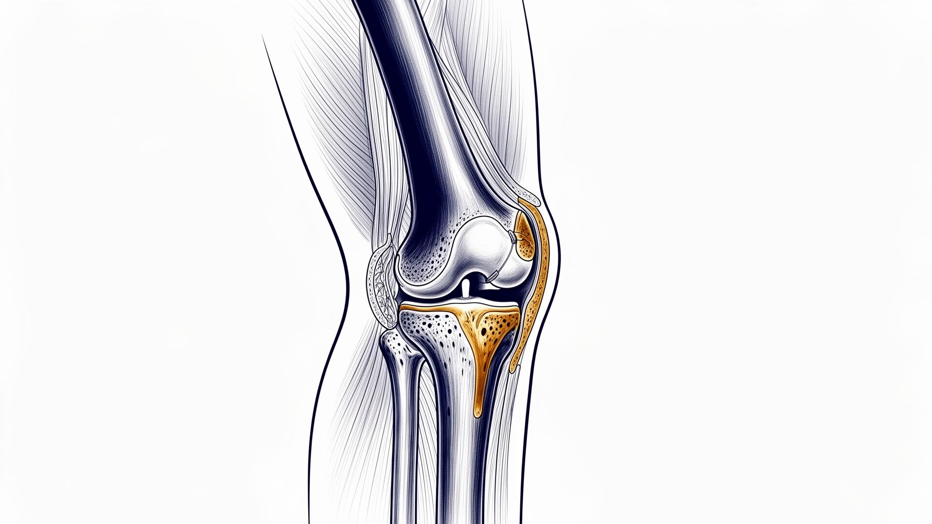
Anatomical illustration of the knee
Treatment areas

Knee surgery

Prosthetic and arthroscopic surgery, corrective osteotomies. Precision techniques for degenerative and sports-related knee conditions.

Introduction

A complex joint, a tailored approach

The knee is one of the most complex and heavily loaded joints in the human body. It bears body weight, enables walking and allows precise movement during sports activity. When osteoarthritis, trauma or degenerative disease impair its function, a tailored surgical approach supported by the most modern technologies becomes essential.

Prof. Simone Cerciello treats the full range of knee pathology: from prosthetic surgery with traditional, robot-assisted and computer-navigated techniques, to minimally invasive arthroscopic surgery for meniscal, ligament and patellar injuries, and corrective osteotomies to preserve the joint in younger patients.

01 · Prosthetic surgery

Knee replacement

Total or partial replacement of the joint with state-of-the-art implants.

Knee replacement is indicated when joint pain and functional impairment, most often caused by advanced osteoarthritis, no longer respond to conservative treatment (medication, injections, physiotherapy).

The procedure replaces the damaged articular surfaces with prosthetic components made of metal alloy (chromium-cobalt or titanium) and highly cross-linked polyethylene. Modern prostheses are designed to faithfully reproduce the natural kinematics of the knee and provide an average lifespan of more than 15-20 years.

The choice between total and unicompartmental replacement — and whether to use robotic or navigation technology — is defined on an individual basis, according to the patient's age, degree of joint degeneration, limb alignment and functional expectations.

With or without navigation / robot

Traditional prosthetic surgery

The classic surgical approach relies on the surgeon's experience and on mechanical instrumentation for the positioning of prosthetic components. In experienced hands this technique delivers excellent results and still represents the reference standard for many patients.

Indications: regular anatomy, standard cases, patients with good bone quality.

Traditional

Computer-navigated surgery

Computer navigation uses an optical tracking system that provides the surgeon with real-time information on limb alignment and component positioning, without altering the surgical gesture.

It reduces variability and yields more accurate alignment, particularly useful in cases with deformity or complex anatomy.

Advantages: greater alignment accuracy, fewer outliers, intra-operative control of ligament balance.

Computer-assisted

Robotic surgery

Latest-generation robotic platforms integrate personalised pre-operative planning based on the patient's CT or X-rays with a robotic arm that assists the surgeon during bone cuts with millimetric precision.

The surgeon always retains control of the procedure: the robot is a precision instrument, not an autonomous executor. Particularly indicated in younger patients, in complex cases and in unicompartmental prostheses.

Advantages: personalised 3D planning, sub-millimetric accuracy, potentially faster recovery, soft-tissue preservation.

Robot-assisted

Total knee replacement

What it is

Total knee replacement (TKR) resurfaces all three articular compartments: the femoral condyle (lower end of the femur), the tibial plateau (upper end of the tibia) and, when needed, the posterior surface of the patella. A highly cross-linked polyethylene insert is placed between the metal components as a bearing surface.

When to operate

The main indication is advanced tricompartmental osteoarthritis with disabling pain, significant functional limitation and failure of conservative treatment.

  • Rheumatoid and other inflammatory arthritis
  • Complex intra-articular fracture sequelae
  • Extensive avascular necrosis of the condyle
  • Severe varus or valgus deformity

How it is performed

The procedure, lasting on average 60-90 minutes, is carried out under spinal or general anaesthesia. Through an anterior incision the surgeon accesses the joint, removes the worn cartilage surfaces and implants the prosthetic components, cemented or press-fit depending on the clinical case.

Recovery

Standing and the first steps take place the day after surgery, with crutches. Average hospital stay is 3-5 days. The rehabilitation programme, lasting 2-3 months, includes mobilisation, muscle strengthening and gait re-education. Return to normal daily activities is usually achieved within 6-8 weeks; complete functional recovery is reached between the third and sixth month.

Unicompartmental knee replacement

What it is

Unicompartmental knee arthroplasty (UKA) is a partial replacement that resurfaces only one compartment of the joint: most often the medial compartment, less commonly the lateral or the patellofemoral. The other articular surfaces, the remaining menisci and both cruciate ligaments are preserved.

When to operate

The procedure is indicated in patients with osteoarthritis isolated and well localised to a single compartment, provided:

  • Intact and functional anterior cruciate ligament
  • Limited deformity (passively correctable varus or valgus)
  • Adjacent compartments in good condition
  • Good pre-operative range of motion
  • Body mass index within acceptable limits

Advantages over total replacement

  • Minimally invasive approach with smaller incision
  • Preservation of bone, healthy cartilage, menisci and ligaments
  • Better proprioception and more physiological kinematics
  • Faster recovery: early walking and quicker return to activities
  • Generally superior post-operative range of motion
  • Easier conversion to total replacement if required in the future

Recovery

Standing is possible on the same day or the day after surgery. Average hospital stay is 2-3 days. Most patients return to normal daily activities within 3-4 weeks, with a complete rehabilitation programme in 6-8 weeks.

02 · Arthroscopic surgery

Knee arthroscopy

Minimally invasive precision techniques for meniscal, ligament and patellar injuries.

Arthroscopy is a minimally invasive technique that allows the surgeon to explore and treat internal knee structures through small incisions (usually two or three, about 5 mm each). A high-definition micro-camera (the arthroscope) is introduced into the joint and projects magnified images onto a monitor, while surgical instruments are introduced through the other incisions.

Compared with traditional open surgery, arthroscopy offers reduced surgical trauma, faster recovery, less post-operative pain and minimal scarring. It is performed as a day-case or with a short stay, most often under spinal anaesthesia.

Meniscal tears

What the meniscus is

The menisci are two fibrocartilaginous crescent-shaped structures (one medial and one lateral) interposed between the femur and the tibia. They absorb loads, distribute forces on the joint, contribute to knee stability and to joint lubrication.

Types of tears

  • Traumatic tears: typical of young patients and athletes, caused by twisting of the knee under load. The most common patterns are longitudinal, radial and 'bucket handle' tears.
  • Degenerative tears: more common after the age of 40-50, related to wear of the meniscal tissue and often associated with early osteoarthritis.

Typical symptoms

Localised pain along the joint line (medial or lateral), swelling, sensation of locking or clicking during movement, difficulty in full extension, pain in squatting and twisting.

Diagnosis

Diagnosis is based on the specialist visit (clinical tests such as McMurray, Apley, Thessaly) and on MRI, which allows accurate assessment of the location, extent and morphology of the tear.

Surgical treatment

Treatment is chosen based on the patient's age, the type and location of the tear and the quality of the remaining tissue:

  • Meniscal repair: first choice whenever possible, especially in younger patients and in tears of the vascularised (red) zone.
  • Selective meniscectomy: targeted removal of the torn portion only, preserving as much healthy tissue as possible.
  • Meniscal transplant: reserved for selected young patients with extensive meniscal loss.

Recovery

After a meniscal repair, weight-bearing is progressively resumed over 4-6 weeks. After a selective meniscectomy, weight-bearing is usually immediate and return to activities is possible within 3-4 weeks.

Anterior cruciate ligament (ACL) injuries

What the ACL is

The anterior cruciate ligament (ACL) is one of the four main ligamentous structures of the knee. It extends from the femoral intercondylar area to the front of the tibial plateau and plays a fundamental role in anterior and rotational stability, especially during pivoting, cutting and deceleration.

Injury mechanism

ACL rupture typically occurs during sports involving changes of direction (football, skiing, basketball, volleyball, rugby, tennis). The most common mechanism is twisting of the knee with the foot fixed to the ground, often without direct contact. The patient hears a 'pop' followed by rapid swelling and a feeling of instability.

Diagnosis

Clinical examination with specific tests (Lachman, pivot shift, anterior drawer) suggests the diagnosis. MRI confirms the diagnosis and assesses any associated injuries (menisci, cartilage, other ligaments).

Treatment

The choice between conservative and surgical treatment depends on the patient's age and activity level, the sport practised, functional instability and associated injuries. In young, active or unstable patients, arthroscopic reconstruction is indicated: the torn ligament is replaced with an autologous graft taken from the patient.

  • Semitendinosus-gracilis (ST-G): harvesting of the pes anserinus tendons, currently the most widespread technique
  • Bone-patellar tendon-bone (BPTB): harvesting of a central portion of the patellar tendon with bone blocks
  • Quadriceps tendon: valid alternative with good functional outcomes

Recovery

Rehabilitation after ACL reconstruction follows a progressive protocol:

  • 0-2 weeks: pain and swelling control, early mobilisation
  • 2-6 weeks: recovery of full range of motion
  • 6 weeks - 3 months: progressive muscle strengthening, proprioception
  • 3-6 months: sport-specific retraining
  • 6-9 months: gradual return to competitive activity after functional and isokinetic testing

Patellar dislocations

What patellar dislocation is

Patellar dislocation is a displacement of the kneecap from its natural seat (the femoral trochlea), almost always laterally. It may occur as an isolated traumatic event or become recurrent in patients with predisposing anatomical factors.

Predisposing factors

  • Trochlear dysplasia: shallow femoral trochlea
  • Patella alta
  • Increased TT-TG distance (tibial tubercle to trochlear groove)
  • Valgus knee or external tibial rotation
  • Generalised ligament laxity
  • Insufficiency of the medial patellofemoral ligament (MPFL), injured during the first dislocation

Symptoms

Acute pain, feeling of the knee 'giving way', swelling, difficulty extending the knee. In recurrent forms, repeated instability even during common activities.

Diagnosis

The specialist visit is complemented by dedicated imaging:

  • Radiographs (axial patellar view at 30°, weight-bearing views)
  • MRI to evaluate cartilage, MPFL and trochlear morphology
  • CT scan with TT-TG measurement in complex cases

Treatment

The first episode is usually treated conservatively: reduction, temporary immobilisation and targeted rehabilitation focused on quadriceps strengthening and motor control. In recurrent dislocations or patients with marked predisposing factors, personalised ('à la carte') surgical treatment is indicated:

  • Reconstruction of the medial patellofemoral ligament (MPFL)
  • Tibial tubercle transposition (Elmslie-Trillat or Fulkerson osteotomy)
  • Trochleoplasty: remodelling of the trochlea in severe dysplasia
  • Lateral release: release of tight lateral structures

Recovery

After MPFL reconstruction, weight-bearing is progressive in the first 2-3 weeks. Return to sport usually occurs 4-6 months later.

03 · Osteotomies

Corrective knee osteotomies

Joint-preserving surgery to realign the limb and delay a prosthesis.

What an osteotomy is

An osteotomy is a joint-preserving procedure that corrects the alignment of the lower limb by modifying the orientation of the bones that form the knee (tibia or femur). The goal is to shift load from the worn compartment to the healthy compartment, reducing pain and significantly slowing the progression of osteoarthritis.

Who it is indicated for

Osteotomy is particularly indicated in young or middle-aged patients (typically 40-60 years) with:

  • Unicompartmental knee osteoarthritis (medial in varus knees, lateral in valgus knees)
  • Limb malalignment (varus or valgus)
  • Joint still well preserved in the other compartments
  • Good ligament stability
  • Desire to maintain an active lifestyle and delay a possible prosthesis

Main types

  • High tibial osteotomy (HTO): indicated in varus knees with medial osteoarthritis. The most common technique is a medial opening wedge, stabilised with a dedicated plate.
  • Distal femoral osteotomy (DFO): indicated in valgus knees with lateral osteoarthritis. Correction is performed on the distal femur.
  • Tibial tubercle transposition osteotomy: used in patellofemoral disorders to realign the patellar traction vector.

Technique and planning

The procedure requires accurate pre-operative planning: full-length weight-bearing radiographs allow precise calculation of the correction needed. Modern technology also enables personalised 3D planning and the use of patient-specific 3D-printed cutting guides.

Recovery

  • Partial or touch-down weight-bearing with crutches for 4-6 weeks
  • Early knee mobilisation from the first days
  • Full weight-bearing at 6-8 weeks, based on radiographic consolidation
  • Return to low-impact sports at 3-4 months, to more demanding activities at 5-6 months
Specialist evaluation

A specialist evaluation for your knee

Every patient is unique. The choice of treatment — conservative, arthroscopic, osteotomy or replacement — is defined after an accurate clinical assessment, supported by the appropriate imaging and tailored to your functional needs and lifestyle.

Mon-Fri · 4:00 pm - 6:00 pm

The information on this page is intended for general educational purposes and does not replace a specialist visit or the physician's advice. Any surgical indication is defined exclusively after a direct clinical evaluation and the appropriate imaging. Content drafted in accordance with the FNOMCeO guidelines (arts. 55-56-57 of the Italian Medical Code of Ethics).